

Working in the field of medical imaging and visualization often produces
images which show occasionally a certain kind of visual beauty. However,
keep in mind that this data is always associated with some form of disease !
The purpose of this page is to display the results of my scientific work
and explain a little bit of its background.
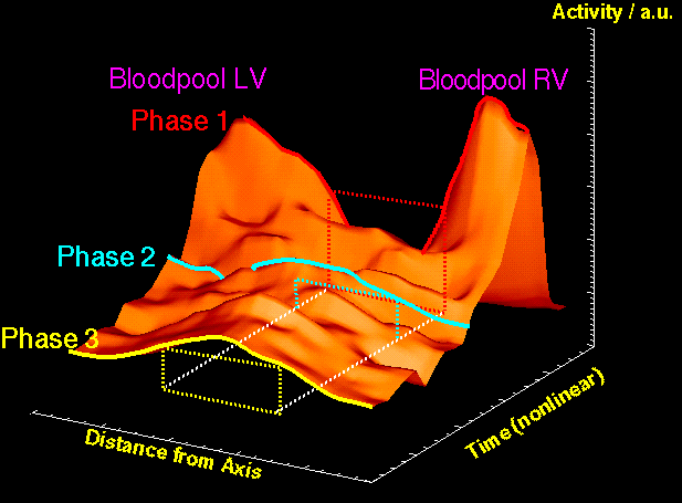
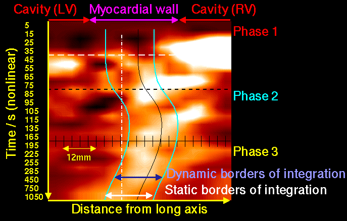
Using N-13 labelled ammonia in PET cardiac imaging, one is able to quantitively assess myocardial flow. The images left show the distribution of ammonia as a function of space (along a line from the center of the hearts's left ventricle through the myocardial septal wall, along the x-axis) and time (after application in a vene, along the y-axis). The concentration of ammonia is displayed as image intensity (left) and height (along the z-axis, right image). As the mentioned line passes through the left and the right ventricle, one can appreciate the distribution of ammonia through the heart in thress phases: 1.) from the vene, it flows through the right ventricle to the lungs 2.) with a small delay it shows up in the left ventricle and finally 3.) it accumulates in the myocardial wall. Of interest is , that the patient's heart (so so did the patient...) moved. Thus, it was the purpose of the study to detect that motion, extract the appropriate concentrations and use those values the perform a pharmaco- kinetical modelling to compute the absolute myocardial blood flow. ( see also).
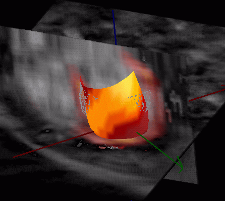

Gated cardiac PET studies allow the simultaneous assessment of FDG uptake , a radiopharmaceutical which resembles the sugar consumption in the heart and cardiac wall motion. The images show the a patient study with an inferolateral infarct (left: enddiastolic i.e. relaxed, right: endsystolic i.e. during maximal compression). As both a gated PET and a gated MRI study was performed (the latter is considered to be the "gold standard" when assessing wall motion) , we are able to visualize the coregistered data sets. This is necessary, as due to the different imaging techniques, a validation is necessary when extracting wall motion from PET data. In addition to the display of orthogonal planes with the fused PET and MRI information, the endocardial border of the left ventricle is shown as surface. Mapped onto this surface is the endocardial shortening which measures the wall motion and thus the ability to pump blood (yellow: high, red: low). Note, that the red areas precisely coincide with a reduced uptake of FDG indicating a reduced metabolism in those regions.

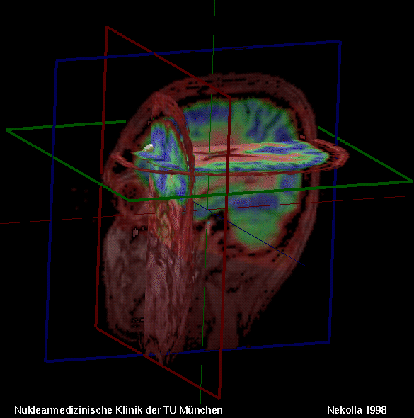 Image coregistration and fusion is a very important field in medical imaging.
Beyond a "show" effect, it is of high relevance to visualize data from
different sources in one image and provide good means of interactivity.
The examples on the left side shows a combination of a FDG PET image and
the corresponding MRI data. The MRI data was used to model the surface of the
skin (grey) while the PET data was used to extract a 3D model of a brain tumor
(hot metal) and display a tilted plane with FDG PET data mapped onto it. The
rendering was done with IDL 4.x and took more than 5 seconds per view.
Using IDL 5.x ObjectGraphics, the right example can be rendered in les than
100 milliseconds allowing full interaction. It displays in the orthogonal
cross sections fused MRI (red/gray) and FMZ PET (prism) data. In the point
where all images coincide, a surface was rendered manually from a comparison
of FMZ PET data with a data base build from normal distribution of FMZ uptake
in the brain. It closely corresponds to a region in this patient's brain
where a epilepsy focus could be located.
Image coregistration and fusion is a very important field in medical imaging.
Beyond a "show" effect, it is of high relevance to visualize data from
different sources in one image and provide good means of interactivity.
The examples on the left side shows a combination of a FDG PET image and
the corresponding MRI data. The MRI data was used to model the surface of the
skin (grey) while the PET data was used to extract a 3D model of a brain tumor
(hot metal) and display a tilted plane with FDG PET data mapped onto it. The
rendering was done with IDL 4.x and took more than 5 seconds per view.
Using IDL 5.x ObjectGraphics, the right example can be rendered in les than
100 milliseconds allowing full interaction. It displays in the orthogonal
cross sections fused MRI (red/gray) and FMZ PET (prism) data. In the point
where all images coincide, a surface was rendered manually from a comparison
of FMZ PET data with a data base build from normal distribution of FMZ uptake
in the brain. It closely corresponds to a region in this patient's brain
where a epilepsy focus could be located.
[Research] [Private] [Gallery] [MunichHeart] [Resources] [MRI|NM]
© Stephan Nekolla 2001